Dental health check

The first important step to your new denture is to make an exact diagnosis as part of a consultation. It begins by filling out an anamnesis form so that the dentist can get important information regarding any illnesses, medical use or other factors that can influence dental treatment.
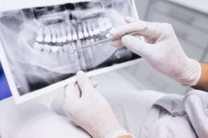
Then a two-dimensional digital panoramic X-ray is taken, which provides the treating dentist with important information about the status, i.e. the condition of the existing teeth, about any missing teeth, misaligned teeth, inflammation and the bone situation. Based on this, pretty precise treatment plans can be drawn up and discussed with the patient. Since there can usually be several ways to correct a tooth problem, it is very important in this phase that the patient is fully informed about the condition and the possible solutions, because the decision about which dental prosthesis is made affects life afterwards. Against this background, it goes without saying that the doctors take enough time for this important preliminary examination, because the patient must answer all questions, show all possible solutions, be informed about advantages and disadvantages, so that he / she can make a responsible decision.
Digital panoramic X-ray
As a first step, we make a digital panorama X-rayConsultation
In the second step we discuss your problems and hear what you were looking for.Examination
In the third step, the dentist will examine you.Treatment plan
At the end of your visit, we will discuss a treatment plan that you will receive from us.The aim of the consultation is that the dentist and patient get a perfect overview of the situation and its possible solutions. With certain types of treatment, further image designs may be required here, e.g. a small x-ray if the canals of certain teeth are checked, a tele-x-ray for an Invisalign treatment or a Cone Beam CT scan before surgery.
When is a CT scan needed?
Before surgery, such as an implant surgery, preliminary examinations must be carried out. The basis is the panoramic x-ray, which is used to evaluate which teeth need to be replaced. Then it has to be assessed whether there is enough bone mass to be able to place implants, or whether bone augmentation or a sinus lift is needed first or simultaneously. Since the panorama X-ray is a two-dimensional image that only shows height and width, a 3D image with a CT or volume tomograph must also be taken to check the bone depth. Furthermore, only a CT scan allows an exact measurement of the amount of bone, on which the size of the implants to be used depends. In addition to bone mass, the general state of health of the patient also plays a crucial role in assessing whether appropriate treatment is possible.


